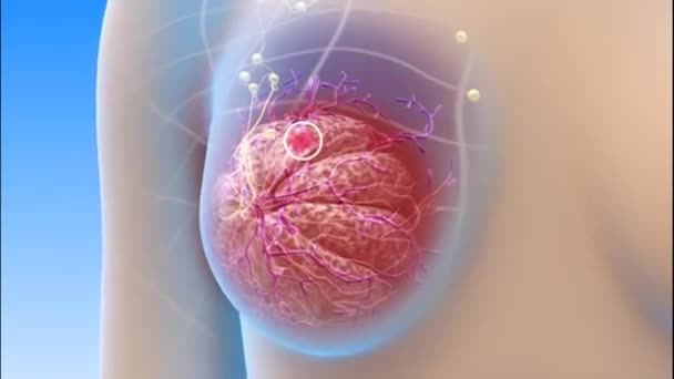
Breast ultrasound has become an essential imaging modality in the early detection and diagnosis of breast diseases. It plays a vital role in evaluating breast lumps, differentiating between cystic and solid lesions, and guiding minimally invasive procedures such as biopsies. At Pink Ribbon Hospital, breast ultrasound is performed using advanced imaging technology to ensure accurate, safe, and patient-centered care for women of all ages.
Unlike mammography, ultrasound does not use ionizing radiation, making it a safe imaging technique, particularly for younger women, pregnant patients, and individuals requiring repeated follow-up examinations. Breast ultrasound is especially beneficial in women with dense breast tissue, where mammography findings may sometimes be limited. The modality provides real-time imaging, allowing radiologists to assess blood flow, tissue characteristics, and lesion morphology with high precision.
Breast ultrasound is frequently used as a complementary examination alongside mammography. When combined, these modalities significantly improve diagnostic confidence and assist clinicians in the early detection of breast cancer. Early diagnosis remains the cornerstone of successful treatment, reducing morbidity and improving survival outcomes for patients.
At Pink Ribbon Hospital, breast imaging services are strengthened by the use of the GE LOGIQ P7 Ultrasound System, an advanced ultrasound platform designed to provide exceptional image quality, workflow efficiency, and patient comfort.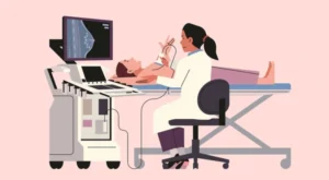
Key Specifications and Features of the GE LOGIQ P7 Ultrasound System
The GE LOGIQ P7 Ultrasound System is recognized for its versatility and high-performance imaging capabilities in breast and general ultrasound applications. Important specifications and features include
- High-resolution imaging technology for improved visualization of breast tissue and small lesions.
- Advanced Doppler imaging for evaluating vascularity and blood flow within breast abnormalities.
- HD Color and B-Flow imaging technologies for enhanced visualization of small vessels and slow blood flow.
- Contrast imaging capability with excellent tissue suppression and image uniformity for enhanced diagnostic confidence.
- Elastography support, including 2D Shear Wave technology, which assists in assessing tissue stiffness and differentiating benign from suspicious lesions.