Breast Cancer is increasing at an alarming rate in Pakistan. 1in 9 women is prone to having breast cancer sometime in her life. This is a worrisome situation that calls for active efforts for screening this disease and nipping the evil in its bud.
Mammography and Ultrasound are the two most widely used imaging modalities for workup of a breast lesion. Both have different indications depending upon patient’s age, type of breast, risk factors and symptoms.
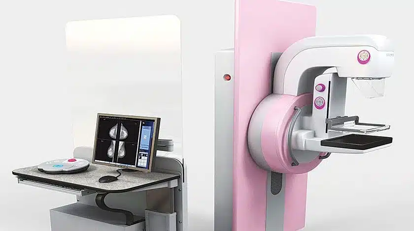
A mammography uses low-dose X-ray to produce breast images known as a mammogram
Mammogram is one of the most commonly used imaging modality for early detection and screening of breast cancer. That means it can also give most information in women who do not have any breast symptoms. It is highly effective in detecting calcifications, distortions, asymmetry in the breast. It is also not dependent on operator so accuracy is not dependent on the expertise of the operator. But mammogram isn’t the ideal tool to detect lesions in women with dense and glandular breasts. Small lesions and tumors are likely to be missed in such breast tissue which is why it is mostly used in women above 40 years of age when the breast tissue mostly becomes fatty. On account of reducing these limitations, Pink Ribbon Hospital is introducing advanced 50 micron 3D mammogram, first of its kind in Pakistan, which produces a 3D image of breast as compared to regular mammograms which produce 2D image, picks smaller lesions with sharper imaging and lesser radiation exposure.
The decision about the kind of imaging modality you need depends on your age, type of breast, symptoms and risk factors and should be taken by your doctor.
An ultrasound uses high-frequency sound waves on the breast and converts them into images. A breast ultrasound is generally used for diagnostic reasons such as when a lump is palpated in clinical examination or when a mammogram reveals suspicious density. Since ultrasound is the recommended imaging modality in women with dense and glandular breast, it is mostly used as first line of investigation in women under forty years of age or in older women with dense parenchyma. One limitation of ultrasound is that it is operator dependent and its accuracy depends on the expertise of the person doing the ultrasound. It also is unable to give image of the entire breasts at once hence not applicable for screening purposes.
Most of the times, ultrasound is used as a supplemental test with mammogram in workup of a lump because it has an ability to differentiate between a solid mass and a fluid filled cyst which then can be further investigated on Fine Needle Aspiration Cytology.
Both the Imaging modalities have their own advantages and limitations and are used as deemed appropriate by a doctor.
WhatsApp us