
A breast ultrasound/mammogram is most often done to find out if a problem found on physical exam of the breast may be a tumor or not. Imaging is very crucial in early diagnosis of breast cancer therefore you should never hesitate to go for it even if just to be sure because there is no harm in it.
AGE: Women younger than 35 are most often advised breast ultrasound because of high breast density but mammo may be used in certain cases as advised by your GP.
Similarly women older than 35 are advised breast mammogram BUT they might have to go through ultrasound as well in case of any ambiguous findings.
Is it painful?
The procedures are not painful at all unless you already have some pain in your breast. In that case you can conveniently tell the doctor to be careful around the area of pain.
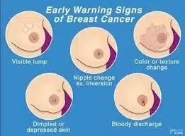
A lot of changes in your breast can indicate the need for immediate imagining.
Lump:
It is important to remember that all lumps are not cancerous. If u find a lump in your breast then you should consider imaging if.
You have family history of breast cancer( very crucial)
The lump is hard or rough
The lump is fixed in position or warmer to touch
There are skin changes around the area of lump.
Any lumps in your armpit
In case your lump is soft, freely mobile and there are no other signs then you need not panic it is most probably benign (curable/not harmful) but you should still get it evaluated by your doctor specially if it starts to grow or you observe any other changes.
Nipple Changes:
Consider imaging if your nipple shows
Bloody Discharge:
Nipple retraction (turning inward)
Slit like nipple (like straight a straight line)
Itching or skin erosion around nipple or areola
Skin Changes:
Erosions or ulcers on skin of breast
Orange peel like appearance of breast
Warmer or reddish skin of the breast than rest of the body
Skin itching or dimpling

What Happens In Breast Imaging
Ultrasound
You will be asked to remove any jewelry and clothing from the waist up. You will be given a gown to wear.
You will lie on your back on an exam table. You will be asked to raise your arm above your head on the side of the breast to be looked at. Or you may be placed on your side.
The technologist will put a clear, warm gel on the skin over the breast area to be looked at.
The technologist will press the transducer against the skin and move it over the area being studied.
Once the test is done, the technologist will wipe off the gel.
You do not need to stop eating or drinking before the test. You also will not need medicine to help you relax.
Mammogram
You will stand in front of a special X-ray machine. A technologist will place your breast on a plastic plate.
Another plate will firmly press your breast from above. The plates will flatten the breast, holding it still while the X-ray is being taken.
You will feel some pressure. The steps are repeated to make a side view of the breast.
The other breast will be X-rayed in the same way.
What’ Next?
• If your imaging tests reveal no suspicious signs then you don’t have to undergo any further tests. Your doctor can give you medicine for any bothering symptom otherwise you are good to go. For future you should continue self-examination and report any changes that you may notice.
• If your imaging tests show any suspicious findings you will have to undergo further tests like biopsy, CT scan and bone scan to confirm your diagnosis and plan your treatment.
But!
You need to remember that diagnosis of breast cancer is not a death sentence. Early diagnosis gives you 93% chances of survival and a healthy life. You need to keep your will strong and get yourself tested as soon as you find a problem.
WhatsApp us